According to the CDC, almost 24 million Americans have diabetes, and it is estimated that six million of those individuals are undiagnosed.1 In 2007, financial costs attributed to diabetes totaled $174 billion.2 Additionally, comorbidities linked with diabetes may lead to serious complications and create additional economic and individual burdens. The development of diabetic foot ulcers (DFUs) is one such complication.
DFU treatment utilizes a considerable portion of health-care dollars and may also lead to significant disability and a decrease in quality of life. Patients with diabetes have a 15%-25% lifetime risk for developing a foot ulcer.3,4 When ulceration occurs, the risk for infection is present and may range in severity from a superficial area to one that pervades the bone. About 25% of diabetic foot infections will extend to deeper subcutaneous tissue or bone, and up to 50% of those individuals will have a recurrent ulcer within the next few years.5 Infection is the leading risk factor for amputation among those with DFUs.6
Considering the prevalence of diabetes, it is conceivable that most primary-care providers (PCPs) will encounter patients with foot ulcers. Practitioners treating patients with diabetes must focus on prevention of ulcerations, prompt diagnosis, treatment initiation, and appropriate referrals to preserve optimal functioning. Given the number of uninsured or underinsured individuals, adhering to this seemingly straightforward strategy can prove difficult. Lack of access to primary care leads many patients to delay care, which prevents detection or delays diagnosis until the advanced stages of disease.
The following case illustrates the challenges faced by many PCPs when caring for an underinsured patient with type 2 diabetes mellitus (DM) and a DFU.CASE STUDY
Mr. J, aged 45 years, was hospitalized for two weeks with cellulitis and a left plantar foot ulcer. Incision and drainage (I & D) revealed three purulent sinus tracts extending from the superficial to the deep space of the left foot involving the first and second metatarsal heads. An MRI established the presence of osteomyelitis, but fortunately the bone was viable, and amputation was avoided. Culture was positive for group B Streptococcus and methicillin-susceptible Staphylococcus aureus, and the patient was treated with antibiotics for six weeks. Ankle-brachial indexes (ABIs) and toe waveforms were within normal limits. On admission, Mr. J's blood sugar was 312 mg/dL and hemoglobin (Hb) A1c was 12.9%. He was diagnosed with type 2 DM and started on insulin therapy. Mr. J achieved good glycemic control, extensive diabetes education was provided, and he was discharged to follow-up in the primary-care setting.
POST-HOSPITALIZATION FOLLOW-UP
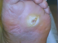 Over the next six months, Mr. J lost 83 lbs, his HbA1c dropped to 5.5%, and his foot ulcer healed. He kept regular appointments with a podiatrist and maintained routine foot care. A small blister was discovered near the site of the previous left plantar DFU (Figure 1). Antibiotic therapy was ordered along with an OTC antimicrobial ointment to be applied to the wound site. While the culture showed no infection and x-ray did not reveal osteomyelitis, the ulcer continued to extrude a small amount of nonodorous serosanguinous drainage. After caring for Mr. J for two months, the PCP referred him to a hospital outpatient wound-care clinic.
No comments:
Post a Comment